Categories
Amniocentesis Test blog page Doppler Test Fetal Medicine Nutrition Pregnancy Test

Why Can’t Parents Hear the Fetal Heartbeat in Early Pregnancy Even When Doctors Confirm It?

One of the most anticipated and joyous moments during early pregnancy is hearing the fetal heartbeat. However, many parents experience confusion or anxiety when their doctor confirms a fetal heartbeat on an ultrasound, yet they themselves cannot hear it. Understanding why this happens can provide reassurance and clarity during those first weeks.

Why Can’t Parents Hear the Fetal Heartbeat Early On?

There are several reasons why parents may not be able to hear the heartbeat even though it is detected by medical equipment:

  • Ultrasound Imaging vs. Doppler Device:
    Doctors often use ultrasound scans (either transvaginal or abdominal) during early pregnancy to visualize the fetal heartbeat. The heartbeat is seen as a flicker or motion on the screen. The “sound” heard during these scans usually comes from the ultrasound machine’s speaker and is not the actual heartbeat sound audible to parents.
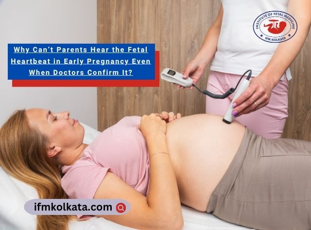
    In contrast, handheld Doppler devices, which pick up sound waves from the heartbeat, are typically used later in pregnancy to allow parents to hear the heartbeat directly.
  • Early Pregnancy Limitations:
    Before approximately 10 to 12 weeks of gestation, the fetal heart is extremely small, and the sound waves it generates are very faint. This makes it difficult for Doppler devices to pick up and amplify the heartbeat sound reliably.
  • Position and Size of the Fetus:
    The fetus’s position inside the uterus and its small size can also affect the clarity and audibility of the heartbeat sound. If the baby is positioned deep inside the uterus or behind the placenta, it may be harder to detect the heartbeat audibly.

When Can Parents Expect to Hear the Fetal Heartbeat?

  • Typical Timeline for Audible Heartbeat:
    Most parents can hear the fetal heartbeat using a Doppler device or a stethoscope-like tool around 10 to 12 weeks of pregnancy. This is when the fetal heart has grown enough and the sound waves are strong enough for detection.
  • Before Audible Detection:
    Prior to this period, doctors primarily rely on ultrasound imaging to confirm the heartbeat visually rather than audibly.

Tips for Parents During Early Pregnancy

  • Don’t Panic if You Can’t Hear It:
    Not hearing the heartbeat at an early stage is completely normal and usually not a sign of any problem.
  • Trust the Doctor’s Ultrasound Confirmation:
    The visual confirmation of a heartbeat via ultrasound is the most reliable and important sign of fetal viability.
  • Ask Questions and Stay Informed:
    Use your prenatal visits to clarify doubts and ask your healthcare provider for reassurance and explanation.
  • Be Patient:
    Remember that fetal development and detection technology have limits in very early pregnancy, and the audible heartbeat is usually detected soon after this early phase.

Hearing the fetal heartbeat is an exciting milestone, but parents should not worry if it is not audible in the very early weeks. The doctor’s confirmation through ultrasound is what truly matters at this stage. As pregnancy progresses, the heartbeat sound becomes easier to detect and hear, providing increasing reassurance to parents.

Categories
Amniocentesis Test blog page Doppler Test Down Syndrome Fetal Medicine Nutrition Pregnancy Test Thalassemia

Why Transvaginal Scan (TVS) Is Done in Early Pregnancy Over Abdominal Ultrasound (USG)

Early pregnancy is a delicate and crucial period that requires accurate monitoring to ensure the health of both mother and baby. Ultrasound scans play an essential role in confirming pregnancy and assessing early fetal development. Among the different ultrasound methods, Transvaginal Scan (TVS) and Abdominal Ultrasound (USG) are commonly used. However, in the first trimester, TVS is often preferred. This article explores what TVS is, why it is done over abdominal USG in early pregnancy, and its advantages.

What is a Transvaginal Scan (TVS)?

A Transvaginal Scan (TVS) is an ultrasound technique where a small, specially designed probe is gently inserted into the vagina. This approach places the probe closer to the uterus, providing clearer and more detailed images of the early pregnancy structures such as the gestational sac, yolk sac, and fetal pole.

Unlike the abdominal ultrasound (USG), where the probe is moved over the abdomen, TVS bypasses the abdominal wall and bladder, which can obscure or reduce the clarity of early pregnancy images.

Advantages of TVS Over Abdominal Ultrasound in Early Pregnancy

TVS offers several critical advantages during the early weeks of pregnancy, typically up to 12 weeks gestation:

  • Earlier Pregnancy Detection:
    TVS can detect pregnancy as early as 4 to 5 weeks, identifying the gestational sac and fetal pole much earlier than abdominal USG, which usually performs better after 6 weeks.
  • Sharper, Clearer Images:
    Because the probe is closer to the uterus, TVS produces high-resolution images that allow for detailed visualization of early fetal structures, helping in accurate dating and viability assessment.
  • Better Fetal Heartbeat Detection:
    Fetal cardiac activity can be detected earlier and with greater accuracy via TVS, often as early as 5.5 to 6 weeks, which is critical for confirming a viable pregnancy.
  • Early Complication Assessment:
    TVS is the gold standard for diagnosing potential early pregnancy complications such as:

    • Ectopic pregnancy (implantation outside the uterus)
    • Threatened miscarriage
    • Subchorionic haemorrhage
  • Assessment of Uterine and Ovarian Health:
    It allows detailed examination of the uterus, cervix, and ovaries, helping identify fibroids, cysts, or other abnormalities that may affect pregnancy.

When is TVS Recommended?

Your doctor may advise a transvaginal scan in early pregnancy under the following circumstances:

  • Confirmation of Early Pregnancy: To verify pregnancy location and viability.
  • Vaginal Bleeding or Abdominal Pain: To assess risks such as miscarriage or ectopic pregnancy.
  • Inconclusive Abdominal Ultrasound Results: When abdominal USG does not provide clear information.
  • History of Previous Pregnancy Complications: To closely monitor pregnancy development.
  • High-Risk Pregnancies: To ensure early and precise fetal monitoring.

What to Expect During a TVS?

  • The procedure is generally safe and painless.
  • The probe is covered with a sterile sheath and lubricated before insertion.
  • It usually takes 10-20 minutes.
  • No special preparation is required, but a full bladder is generally not needed (unlike abdominal USG).

In early pregnancy, TVS is preferred over abdominal ultrasound due to its superior ability to detect pregnancy earlier, provide clearer images, and identify potential complications promptly. This early and accurate assessment enables better monitoring and management, ensuring the best possible care for mother and baby. If you experience any symptoms or require early pregnancy evaluation, consult your healthcare provider about the benefits of a transvaginal scan.

Categories
Amniocentesis Test blog page Doppler Test Fetal Medicine Pregnancy Test

Viability Scan vs. NT Scan: What’s the Difference?

Understanding Early Pregnancy Scans

Early pregnancy involves several important ultrasound scans to monitor fetal health and development. Two common scans during the first trimester are the viability scan and the nuchal translucency (NT) scan. Although both are ultrasound procedures, they serve different purposes and are performed at different stages.

What is a Viability Scan?

A viability scan is usually performed between 6 to 9 weeks of pregnancy. Its primary goals include:

  • Confirming the presence of an intrauterine pregnancy
  • Detecting a fetal heartbeat
  • Estimating the gestational age of the embryo
  • Checking for multiple pregnancies (twins or more)
  • Identifying early pregnancy complications such as ectopic pregnancy or miscarriage risk

This scan reassures parents that the pregnancy is developing normally in the early stages.

What is the NT Scan?

The Nuchal Translucency (NT) scan is a screening test performed between 11 and 13 weeks of pregnancy. It measures the thickness of the clear fluid-filled space at the back of the baby’s neck called the nuchal translucency.

An increased NT measurement can indicate a higher risk for certain chromosomal abnormalities, including:

  • Down syndrome (Trisomy 21)
  • Edwards syndrome (Trisomy 18)
  • Patau syndrome (Trisomy 13)

The NT scan is usually combined with blood tests to provide a risk assessment for these conditions.

Key Differences Between Viability Scan and NT Scan

Feature

Viability Scan

NT Scan

Timing

6 – 9 weeks

11 – 13 weeks

Purpose

Confirm pregnancy and heartbeat

Screen for chromosomal abnormalities

What it measures

Presence of embryo and heartbeat

Thickness of nuchal translucency

Additional use

Check for multiples, early risks

Combined with blood tests for risk score

Why Are Both Scans Important?

  • The viability scan provides early confirmation and peace of mind that the pregnancy is progressing normally.
  • The NT scan offers valuable information about genetic risks that help parents make informed decisions about further testing and pregnancy management.

Both scans are non-invasive, safe, and vital parts of comprehensive prenatal care.

Understanding the difference between the viability scan and NT scan can help expectant parents navigate early pregnancy with confidence. Always follow your healthcare provider’s advice on when to schedule these important ultrasounds.

Categories
blog page

How Maternal-Fetal Immune System Helps Ensure a Healthy Pregnancy

Pregnancy is a beautiful and complex journey, filled with wonder and anticipation. It’s a time when a mother’s body nurtures and protects her developing baby. But have you ever wondered how the mother’s immune system, responsible for defending against diseases, knows not to attack the growing baby? In this article, we’ll explore the remarkable interplay between the maternal and fetal immune systems, shedding light on how they collaborate to ensure a successful pregnancy.

Basics of Maternal-Fetal Immunity

Pregnancy presents a unique immunological challenge because, under normal circumstances, the mother’s immune system’s primary job is to protect her from infections and foreign invaders. However, during pregnancy, there’s a delicate balancing act that occurs to ensure that the growing baby, which carries genetic material from both parents, is not mistakenly treated as a threat. This challenge is at the heart of maternal-fetal immunology, where nature has devised an incredible system to allow pregnancy to thrive despite these potential complications.

Immunological Challenge: At first glance, it might seem like the baby, carrying half of its genetic material from the father, should be seen as foreign by the mother’s immune system. However, nature has devised a clever system to prevent this.

Placental Barrier: Think of the placenta as a crucial mediator between the mother and the baby. It acts as both a protective barrier and a conduit for essential nutrients and oxygen. This remarkable organ ensures that the mother’s immune system doesn’t view the baby as an intruder.

Immunological Tolerance: The mother’s immune system undergoes a unique adaptation process. It learns to tolerate the presence of the baby’s genetic material and understands that, in this special situation, it should protect the baby rather than launch an immune attack.

Dynamic Interplay

Within the mother’s body, an intricate dance of biological interactions takes place during pregnancy. This dynamic interplay involves various elements of the maternal and fetal immune systems, working in harmony to ensure the well-being of both. It’s a bit like a well-choreographed performance, where different actors—immune cells, messenger molecules, and more—play their roles to perfection. In this section, we’ll delve deeper into this choreography and explore how the immune systems collaborate to create the optimal environment for a healthy pregnancy.

Immune Cells in Pregnancy: Specialized immune cells, including regulatory T cells and macrophages, play key roles in maintaining a harmonious balance between immune tolerance and protection. These cells communicate with the mother’s immune system, ensuring it doesn’t harm the developing baby.

Cytokine Messengers: Cytokines are like messengers in the body, and they play a pivotal role in the communication between the maternal and fetal immune systems. They guide the immune system, telling it when to protect the baby and when to respond to infections.

Challenges and Complications

When Things Go Awry: Sometimes, the delicate balance of the maternal-fetal immune system can be disrupted, leading to complications. Conditions like preeclampsia, which causes high blood pressure in the mother, and fetal growth issues can occur when the immune system doesn’t function as it should.

Infections and Immunology: Infections during pregnancy can alter the immune system’s equilibrium, potentially harming the baby. It’s essential to screen for and manage infections to safeguard both the mother and the baby.

Clinical Implications and Future Directions

Understanding the intricacies of maternal-fetal immunology has not only unlocked the secrets of successful pregnancies but has also led to important clinical applications. One such application involves medical interventions designed to manage and rectify immunological issues when they occur. These interventions can range from immunomodulatory therapies to personalized treatment plans. Beyond the current clinical landscape, ongoing research in this field continues to drive innovation.

Scientists and medical professionals are collaborating to explore novel approaches and cutting-edge techniques that promise to enhance our ability to safeguard the health of both the mother and the growing baby. In this ever-evolving field, the future holds the promise of even more precise and effective strategies for ensuring the well-being of expectant mothers and their little ones.

Medical Interventions: When complications arise due to immune system issues, doctors can step in with medical treatments. These interventions aim to restore the balance in the immune system, ensuring the baby’s safety and well-being.

Continuing Research: Scientists are continually expanding our knowledge of maternal-fetal immunology. Ongoing research seeks to unlock new ways to ensure the safety and health of both the mother and the baby, addressing immunological challenges with greater precision.

Final Words

The interplay between the maternal and fetal immune systems is nothing short of fascinating. It ensures that most pregnancies progress smoothly, culminating in the birth of a healthy baby. When complications arise, the medical community works diligently to find solutions. Understanding this remarkable teamwork enhances our appreciation of the intricate journey of pregnancy and the strides we continue to make in improving the experience for both mother and child.

Categories
blog page Fetal Medicine

How to Choose a Good Fetal Care Clinic for High-Risk Pregnancy

Pregnancy can be risky for some women. The entire process can cause a huge health risk for some would-be moms and their unborn babies too. Thanks to modern medical technologies that nowadays, pregnancy has become less risky than before. If your ob-gyn suspects your case is a high-risk pregnancy, you must opt for a reputable clinic where fetal medicine and care are available.

Kolkata has multiple clinics where Fetal Echocardiography and other clinical tests are done to ensure that the mother and the fetus are in a safe condition. Finding one suitable for you is not tough when you know how to choose the right one.

Vital Factors to Check in a Fetal Care Centre

List of Doctors, Radiologists and Other Technicians

A clinic can provide you with the best service because of its team. Thus, it is important to check the list of doctors and other technicians to ensure that you will receive the best support from the top professionals in this industry. Go through the names of fetal care experts, obstetricians and gynaecologists, along with radiologists and fetal medicine professionals. Check their degrees and experience to be sure about their skills and experience.

Range of Services

A good fetal care unit typically offers a range of services for would-be mothers and their unborn babies. This also includes infertility treatments, special care for high-risk pregnancies, Invasives and Non-invasives, IVF/IUI, USG and blood tests in pregnancy. All these medical examinations are done to monitor the fetus’s and the mom’s health. Fetal medicine experts will check these reports and determine what type of treatments your unborn baby should receive inside your womb to be born healthy.

Advanced Equipment

A modern fetal care unit must have advanced equipment that will help doctors and technicians correct and diagnose the issues in a woman and her unborn baby. The results must be accurate and detect different aspects of one particular medical condition. Complex tests like Fetal Echocardiography and others must be done with modern equipment by skilled and certified
technicians only.

Good Customer Service

Fetal care clinics must have excellent customer service. Generally, people come here with lots of anxiety and stress. It is the responsibility of the clinic’s staff to make them feel comfortable and stress-free with the right guidance and treatments.

IFM Fetal Medicine Unit Kolkata

Institute of Fetal Medicine or IFM offers a range of services and medical options to make pregnancy the most lovable and memorable experience of your life. We have highly advanced equipment in our clinic. Our doctors are the best in their field. Please get in touch with us for more details.