
Categories
Fetal Medicine blog page

Understanding Chromosomal Abnormalities in Pregnancy

Chromosomal abnormalities are genetic conditions that can affect fetal development and are often detected during prenatal screenings. These conditions occur when there is an extra or missing chromosome, or when chromosomes are structurally altered. Understanding chromosomal abnormalities is crucial for expectant parents, as early detection can help in making informed decisions and preparing for the child’s needs post-birth.

What Are Chromosomal Abnormalities?

Chromosomes carry the genetic information necessary for normal development. Humans have 23 pairs of chromosomes, and any change to this number or structure can lead to developmental or health problems. There are several types of chromosomal abnormalities:

  • Trisomy 21 (Down Syndrome): The most common chromosomal abnormality, characterized by an extra 21st chromosome. Children with Down syndrome may experience developmental delays, intellectual disabilities, and potential heart or gastrointestinal problems.
  • Trisomy 18 (Edwards Syndrome): This rare condition involves an extra chromosome 18 and leads to severe developmental delays. Most pregnancies affected by Trisomy 18 end in miscarriage or stillbirth. Those that survive have significant physical and intellectual disabilities.
  • Trisomy 13 (Patau Syndrome): Involves an extra 13th chromosome, causing severe developmental and physical abnormalities. Like Trisomy 18, most pregnancies affected by Trisomy 13 end in miscarriage.

How Are Chromosomal Abnormalities Diagnosed?

Prenatal testing plays a key role in identifying chromosomal abnormalities:

  • Non-Invasive Prenatal Testing (NIPT): This is a blood test that screens for Down syndrome, Trisomy 18, and Trisomy 13. It is non-invasive and highly accurate.
  • Ultrasound: During routine ultrasounds, markers for chromosomal conditions, such as nuchal translucency (a fluid-filled space at the back of the baby’s neck), may be detected.
  • Amniocentesis and CVS (Chorionic Villus Sampling): These invasive tests can provide definitive results by analysing amniotic fluid or placental tissue.

Why Prenatal Genetic Counselling Is Important

When chromosomal abnormalities are suspected or confirmed, genetic counselling becomes essential. It helps parents understand the implications of the findings, discusses the options available, and provides emotional support during this challenging time. Genetic counselling can also help parents prepare for the birth of a child with special needs.

Managing a Pregnancy with Chromosomal Abnormalities

While some chromosomal abnormalities have no known prevention, early detection can help manage the pregnancy and plan for the future. Parents can make decisions regarding additional tests, treatments, or interventions after birth, and healthcare providers can prepare for any specialized care that may be required.


Categories
blog page Fetal Medicine

Recurrent Pregnancy Loss: Causes, Genetic Factors, and Support

Recurrent pregnancy loss (RPL) is a condition where a woman experiences two or more consecutive miscarriages. These losses can occur at any stage of pregnancy, including the early first trimester, second trimester, or, in rare cases, the third trimester. However, RPL is typically defined as miscarriages occurring before 24 weeks of gestation.

Causes of Recurrent Pregnancy Loss

There are several potential causes of RPL, which may vary from case to case:

  1. Uterine Abnormalities: Structural issues with the uterus, such as uterine septum or fibroids, can increase the risk of pregnancy loss.
  2. Infections: Certain infections can impact fetal development, leading to miscarriage.
  3. Thrombophilia: Blood clotting disorders, which can reduce blood flow to the placenta, are another common cause.
  4. Genetic Factors: Chromosomal abnormalities in either parent can result in repeated miscarriages.

Role of Genetic Factors in RPL

Genetic issues contribute to a significant percentage of RPL cases. Chromosomal abnormalities, such as translocations or aneuploidies, may prevent the embryo from developing normally. Genetic counselling and investigations play a critical role in identifying these issues and helping couples understand their reproductive risks.

Additionally, genetic testing may provide insights into neurodevelopmental conditions or other potential health concerns that could affect future pregnancies.

Importance of Genetic Counselling for RPL

Genetic counselling is essential for couples experiencing RPL, as it:

  • Identifies genetic causes of miscarriages.
  • Provides clarity and emotional support to affected couples.
  • Guides them on treatment options or preventative measures, such as preimplantation genetic testing (PGT) during in-vitro fertilization (IVF).

Seeking Expert Support

If you are facing recurrent pregnancy loss or have concerns about your pregnancy, the Institute of Fetal Medicine, Kolkata, offers expert care, genetic counselling, and comprehensive support tailored to your needs.

Contact us:
📞 9830047676 / 9748480005 / 9831788538

Categories
Fetal Medicine blog page

Understanding Renal Problems Diagnosed in the Fetus

Renal or kidney issues can sometimes be diagnosed in a fetus during routine ultrasounds. The kidneys, along with the ureters, bladder, and the entire urinary system, start forming early in fetal development. By around 10 weeks of gestation, these structures are visible on ultrasound. However, anomalies are typically identified during the second-trimester anomaly scan.

Common Fetal Renal Problems

Fetal renal issues can range from mild to severe:

  • Mild Renal Pelvis Dilatation (RPD): A common condition where the renal pelvis, a part of the kidney, is slightly enlarged. This is often due to urine retention or mild obstruction in the passage from the kidney to the bladder.
  • Severe Anomalies: Conditions such as a significantly dilated bladder (megacystis) or complete obstruction of the urinary tract or multicystic dysplastic kidneys.

Renal Pelvis Dilatation (RPD) During the Second Trimester

Renal pelvis dilatation is a condition often detected during the second-trimester anomaly scan. This condition may result from mild urine retention or mechanical obstruction and is usually not a cause for concern.

  • Prognosis: In most cases, RPD resolves on its own during the pregnancy or after birth.
  • Association with Chromosomal Abnormalities: While rare, RPD can be associated with chromosomal issues like Down syndrome. However, the risk is minimal when RPD is an isolated finding without other anomalies.

Importance of Follow-Up Scans

A follow-up scan around 28 weeks is essential to monitor the condition. This reassures parents about the baby’s health and tracks any changes in the urinary system. Most babies diagnosed with mild RPD have a normal outcome after delivery and require little to no medical intervention.

Expert Guidance at the Institute of Fetal Medicine

If you have concerns about your pregnancy or your baby’s health, the Institute of Fetal Medicine, Kolkata, provides expert diagnosis, care, and support for fetal renal problems.

Contact us:
📞 9830047676 / 9748480005 / 9831788538

Categories
blog page Fetal Medicine Nutrition

Foods to Avoid During Pregnancy: Protecting Your Baby’s Health

Understanding which foods to avoid during pregnancy is crucial for ensuring the health of both mother and baby. Some foods can pose significant risks, and being informed can help in making safer choices.

Foods to Avoid

  • Raw or Undercooked Meats: These can harbour harmful bacteria and parasites. Ensure all meats are thoroughly cooked to safe temperatures to minimize risks.
  • High-Mercury Fish: Certain fish, such as shark, swordfish, and king mackerel, can contain high levels of mercury, which is harmful to fetal development. Opt for low-mercury options like salmon, sardines, and shrimp.
  • Unpasteurized Products: Avoid unpasteurized dairy products and juices, which can lead to infections such as Listeria. Stick to pasteurized options to reduce risks.
  • Certain Cheeses: Soft cheeses, such as feta and brie, may pose risks if they are unpasteurized. Always check labels and choose safe options.

Limitations on Caffeine and Alcohol

  • Caffeine: High caffeine intake has been associated with an increased risk of miscarriage and low birth weight. It’s best to limit caffeine consumption to about 200 mg per day, equivalent to one 12-ounce cup of coffee.
  • Alcohol: Pregnant women should avoid alcohol entirely, as it can lead to fetal alcohol spectrum disorders, which can have lifelong effects on the child’s development.

Reading Labels

Being mindful of processed foods and additives can help maintain a healthy diet. Focus on whole, minimally processed foods to ensure better health outcomes for both mother and baby.

Final Words

Being aware of foods to avoid during pregnancy is essential for protecting your baby’s health. By educating yourself about safe food practices and consulting with healthcare providers, expectant mothers can make informed dietary choices that promote a healthier pregnancy.

Categories
blog page Fetal Medicine Nutrition Pregnancy Test

Hydration in Pregnancy: How Much Water Do You Need?

Staying properly hydrated is crucial during pregnancy. Adequate fluid intake supports both maternal and fetal health, helping to maintain overall wellness throughout this important time.

Daily Water Intake Recommendations

Expectant mothers should aim for at least 8-10 cups (64-80 ounces) of fluids daily, adjusting based on individual needs, activity levels, and climate. Staying well-hydrated can prevent many common pregnancy-related issues.

Hydrating Foods

Incorporating hydrating foods into your diet can help meet fluid needs. Fruits and vegetables with high water content, such as watermelon, cucumbers, oranges, and strawberries, not only provide hydration but also essential vitamins and minerals.

Benefits of Proper Hydration

  • Prevents Common Issues: Adequate hydration can help prevent pregnancy-related issues such as constipation, urinary tract infections, and swelling.
  • Supports Amniotic Fluid Levels: Proper hydration is essential for maintaining healthy amniotic fluid levels, which cushions and protects the developing fetus.

Mindful Choices

Choosing water, herbal teas, and other low-sugar beverages can help maintain hydration without adding excessive calories or sugar. Limiting caffeine intake is also advisable, as high caffeine levels can lead to dehydration.

Final Words

Prioritizing hydration is essential for a healthy pregnancy. By making a conscious effort to drink enough water and incorporate hydrating foods into your diet, expectant mothers can support overall health and fetal development effectively.

Categories
blog page Fetal Medicine Nutrition

Nutritional Tips for Managing Gestational Diabetes

Gestational diabetes is a condition that can develop during pregnancy, requiring careful management to ensure the health of both mother and baby. Proper nutrition is essential for regulating blood sugar levels and minimizing risks associated with gestational diabetes.

Key Dietary Strategies

  • Choose Complex Carbohydrates: Opt for whole grains, such as brown rice and whole-wheat bread, which help maintain stable blood sugar levels. These complex carbohydrates are digested more slowly compared to simple sugars, providing a gradual release of energy.
  • Incorporate Lean Protein: Protein helps stabilize blood sugar and keeps you feeling full. Lean proteins such as chicken, turkey, fish, beans, and tofu are excellent choices.
  • Include Healthy Fats: Healthy fats, such as those found in avocados, nuts, and olive oil, can help regulate blood sugar and provide essential nutrients for the developing baby.

Meal Timing

Eating regular meals and snacks throughout the day can help maintain steady glucose levels. It’s beneficial to pair carbohydrates with protein or healthy fats to minimize blood sugar spikes.

Monitor and Consult

Collaborating with healthcare providers, such as a dietitian or nutritionist, can help develop a personalized meal plan tailored to individual needs. Regularly monitoring blood sugar levels is also crucial for effective management.

Final Words

Effectively managing gestational diabetes through nutrition is essential for a healthy pregnancy. By making informed dietary choices and consulting healthcare professionals, expectant mothers can support their health and their baby’s development.

Categories
blog page Fetal Medicine Nutrition

Importance of Prenatal Vitamins for Fetal Development

Prenatal vitamins play a critical role in supporting the health of both expectant mothers and their developing babies. These specialized supplements are designed to fill nutritional gaps and ensure that both the mother and fetus receive the essential nutrients needed for optimal growth and development.

Key Nutrients in Prenatal Vitamins

  • Folic Acid: This B-vitamin is crucial for preventing neural tube defects, which can affect the brain and spine of the fetus. Women are encouraged to start taking folic acid at least a month before conception and continue throughout the first trimester.
  • Iron: Iron is vital for increasing blood volume during pregnancy and preventing anaemia. Pregnant women should aim to consume around 27 mg of iron daily, found in foods like lean meats, spinach, and fortified cereals.
  • Calcium: Necessary for the development of the baby’s bones and teeth, calcium helps maintain the mother’s bone health as well. Pregnant women need about 1,000 mg of calcium per day, which can be obtained from dairy products, leafy greens, and fortified plant-based milks.
  • DHA (Docosahexaenoic Acid): An omega-3 fatty acid important for brain and eye development. Pregnant women should aim for at least 200 mg of DHA daily, primarily found in fatty fish or supplements.

Why Prenatal Vitamins Matter

  • Early Development: The first trimester is a crucial time for fetal development. Adequate folic acid intake is essential during this period to minimize risks associated with neural tube defects.
  • Oxygen and Nutrient Supply: Iron is critical for transporting oxygen to the fetus. Insufficient iron levels can lead to fatigue and complications during delivery.
  • Support for Bone Health: Calcium is not only essential for the baby’s development but also for the mother’s long-term health, especially considering the demands of lactation post-delivery.

Consultation is Key

It’s essential for pregnant women to consult with healthcare providers to determine the right prenatal vitamin tailored to their individual needs. The right dosage and formulation can make a significant difference in ensuring optimal health outcomes.

Final Words

Prioritizing prenatal vitamins is vital for a healthy pregnancy. By educating themselves about essential nutrients and working with healthcare professionals, expectant mothers can enhance their own health and support their baby’s development effectively.

Categories
Amniocentesis Test blog page Fetal Medicine

Amniocentesis: How It Helps in Detecting Genetic Disorders

Amniocentesis is a prenatal diagnostic procedure used to identify genetic disorders and other chromosomal abnormalities in the developing baby. Performed typically between the 15th and 20th weeks of pregnancy, this test involves taking a small sample of the amniotic fluid surrounding the baby in the uterus. This fluid contains fetal cells and various chemicals produced by the baby, which can provide vital information about the baby’s genetic makeup and overall health.

In this blog, we’ll explore how amniocentesis is performed, what conditions it can detect, who should consider it, and the risks involved.

What is Amniocentesis?

Amniocentesis is a diagnostic test that examines the amniotic fluid for chromosomal abnormalities, genetic conditions, and neural tube defects. The test can provide highly accurate results, making it a key tool for detecting conditions like Down syndrome, spina bifida, and cystic fibrosis.

The procedure is considered when there’s an increased risk of genetic abnormalities due to factors such as maternal age, family history, or abnormal results from earlier prenatal screenings.

Who Should Consider Amniocentesis?

Amniocentesis is generally offered to women who are at a higher risk for genetic conditions, including:

  • Women over 35: The risk of chromosomal abnormalities like Down syndrome increases with maternal age.
  • Abnormal screening results: If earlier screening tests (such as first-trimester blood tests or ultrasound) show signs of possible genetic issues, amniocentesis can confirm the diagnosis.
  • Family history of genetic disorders: If there is a known history of certain genetic conditions, amniocentesis can help detect if the baby has inherited them.
  • Previous pregnancy with a genetic condition: Women who have had a previous pregnancy with a genetic disorder may choose amniocentesis in future pregnancies to check for similar conditions.

How is Amniocentesis Performed?

The amniocentesis procedure is typically quick and performed in a medical setting. Here’s how it works:

  1. Ultrasound guidance: An ultrasound is used to determine the baby’s position and locate a safe spot to insert the needle.
  2. Needle insertion: A thin, hollow needle is carefully inserted through the mother’s abdomen and into the uterus. A small sample of the amniotic fluid is drawn into a syringe.
  3. Sample analysis: The collected fluid, which contains fetal cells, is sent to a lab for analysis. These cells are examined for genetic abnormalities and other conditions.

The procedure usually takes about 30 minutes, and most women experience only mild discomfort, similar to a regular blood test.

What Conditions Can Amniocentesis Detect?

Amniocentesis can detect a wide range of genetic and chromosomal conditions, including:

  • Down syndrome (Trisomy 21): A common genetic disorder that affects a baby’s development and intellectual abilities.
  • Spina bifida: A neural tube defect where the spine and spinal cord don’t form properly.
  • Cystic fibrosis: A genetic disorder that affects the lungs and digestive system.
  • Sickle cell anemia: A blood disorder that causes red blood cells to become misshapen.
  • Tay-Sachs disease: A fatal genetic condition that affects the nerve cells in the brain and spinal cord.

It’s important to note that while amniocentesis is highly accurate for detecting these conditions, it does not test for every possible disorder.

Risks of Amniocentesis

Amniocentesis is considered a safe procedure, but like any medical test, it carries some risks. The primary risk is miscarriage, which occurs in about 1 in 200 to 1 in 400 procedures. Other potential risks include:

  • Infection: In rare cases, an infection may occur following the procedure.
  • Rh sensitization: If the mother has Rh-negative blood and the baby is Rh-positive, there’s a risk of Rh sensitization. This can be prevented by receiving a Rh immunoglobulin injection.
  • Vaginal bleeding or leaking amniotic fluid: These side effects are uncommon but can occur after the procedure.

Your healthcare provider will discuss these risks with you to help you make an informed decision about whether amniocentesis is right for you.

Why is Amniocentesis Important?

For many parents, amniocentesis provides crucial insights into the baby’s genetic health. The test offers:

  • Definitive answers: Unlike non-invasive screening tests, amniocentesis provides a clear diagnosis, helping parents understand their baby’s health with confidence.
  • Early preparation: If a genetic condition is detected, parents and healthcare providers can prepare for medical interventions or specialized care that may be required after birth.
  • Informed decision-making: Amniocentesis results can help parents make informed choices about their pregnancy.

Amniocentesis is a valuable diagnostic tool for detecting genetic disorders during pregnancy. While it is an invasive procedure with a small risk of miscarriage, it offers accurate and definitive answers that can help families plan and prepare for their baby’s future health.

If you’re considering amniocentesis, it’s important to consult with your healthcare provider, who can guide you based on your individual risk factors and help you understand the benefits and potential risks of the procedure.

Categories
blog page Fetal Medicine

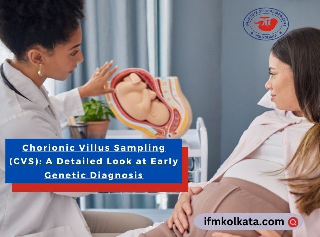
Chorionic Villus Sampling (CVS): Early Genetic Diagnosis

Chorionic Villus Sampling (CVS) is a prenatal test performed between the 10th and 13th weeks of pregnancy to detect genetic abnormalities in the baby. It is particularly useful for diagnosing conditions such as Down syndrome, cystic fibrosis, and other genetic disorders. This test involves taking a small sample of the placenta (chorionic villi) for analysis and is often recommended for women who are at a higher risk of genetic disorders based on age, family history, or abnormal screening results.

In this blog, we will explore how CVS works, who should consider it, the risks involved, and why it is an important diagnostic tool for many parents.

What is Chorionic Villus Sampling (CVS)?

Chorionic Villus Sampling (CVS) is a prenatal test designed to detect certain genetic disorders. During the procedure, a small sample of chorionic villi from the placenta is collected and tested. These cells share the baby’s genetic makeup, allowing doctors to detect chromosomal conditions.

CVS is known for its ability to provide early results, which can be vital for families who want to understand their baby’s health in the first trimester. The test is highly accurate, with results that are almost 99% conclusive for specific conditions.

Who Should Consider CVS?

Not all pregnancies require CVS. It is typically recommended in certain high-risk situations, such as:

  • Maternal Age: Women over the age of 35 are at a higher risk for chromosomal abnormalities like Down syndrome.
  • Family History: If there is a history of genetic disorders, CVS can help detect if the baby has inherited the condition.
  • Abnormal Screening Results: If early blood tests or ultrasounds show signs of potential problems, CVS can confirm or rule out certain conditions.
  • Previous Pregnancy with a Genetic Disorder: If a previous child was born with a genetic disorder, CVS may be recommended in future pregnancies to check for similar issues.

How is the CVS Procedure Performed?

CVS can be performed in two ways: transcervical or transabdominal.

  1. Transcervical CVS: A thin tube is inserted through the cervix to reach the placenta, and a small sample of chorionic villi is removed.
  2. Transabdominal CVS: A needle is inserted through the abdomen into the uterus to collect the sample.

The choice of method depends on the position of the placenta, determined by an ultrasound before the procedure. The procedure itself takes about 30 minutes, though the actual sampling takes only a few minutes.

What Conditions Can CVS Detect?

CVS is primarily used to detect chromosomal abnormalities such as:

  • Down syndrome (Trisomy 21): A condition that causes developmental delays and intellectual disabilities.
  • Cystic fibrosis: A genetic disorder affecting the lungs and digestive system.
  • Sickle cell anemia: A blood disorder that causes red blood cells to become misshapen.
  • Tay-Sachs disease: A fatal genetic disorder that affects nerve cells in the brain and spinal cord.

It is important to note that CVS does not detect all genetic disorders but can identify many severe conditions.

Risks of CVS

CVS is an invasive procedure, and like any medical intervention, it carries some risks. The primary risk is miscarriage, which occurs in about 1 in 100 procedures. Other risks include:

  • Infection: Though rare, there is a small risk of infection following the procedure.
  • Vaginal bleeding: Some women experience light bleeding afterward, which usually resolves on its own.
  • Rh sensitization: If the mother has Rh-negative blood and the baby is Rh-positive, there is a risk of sensitization. However, this can be managed with a Rh immunoglobulin injection.

Why is CVS Important?

CVS provides vital early insights into the baby’s genetic health. For parents facing a higher risk of genetic disorders, this information can be empowering. The test allows families to:

  • Make informed decisions about continuing the pregnancy.
  • Prepare for medical interventions or specialized care that may be needed after birth.
  • Gain peace of mind if the results show no abnormalities.
Categories
blog page Fetal Medicine

Understanding Fetal Ultrasound: What to Expect During Pregnancy Scans

Fetal ultrasound is a key component of prenatal care, providing vital insights into the health and development of the baby during pregnancy. This non-invasive diagnostic procedure uses high-frequency sound waves to create detailed images of the fetus inside the womb. It allows healthcare providers to monitor the growth, detect potential abnormalities, and track the progress of the pregnancy.

Typically, women undergo two main ultrasounds: one during the first trimester to confirm the pregnancy and another between 18 to 20 weeks for a detailed examination of the baby’s anatomy. The second scan, known as the anomaly scan, provides information about the baby’s development, checking for structural abnormalities such as heart defects or neural tube issues. Some women, especially those with high-risk pregnancies, may require additional ultrasounds to ensure the baby is developing as expected.

In addition to tracking growth, fetal ultrasounds can detect the baby’s heartbeat, movements, and even gender if the parents wish to know. This technology has revolutionized prenatal care by offering a non-invasive way to detect potential issues early, allowing doctors and parents to prepare for medical interventions if necessary.

Fetal ultrasounds also provide peace of mind for expectant parents by giving them a visual connection to their developing baby, enhancing the pregnancy experience. These scans are typically safe, with no radiation involved, and pose minimal risk to both mother and baby.

Fetal ultrasound plays a crucial role in prenatal care, ensuring that both the mother and the healthcare team are informed about the baby’s well-being throughout the pregnancy. Early detection of potential issues can lead to better outcomes and more personalized care, helping ensure a healthy and safe pregnancy for both mother and baby.