Hướng Dẫn Cách Nhận Freespin Hằng Tuần Khi Chơi Nổ Hũ Tại SHBET | shbetvy.com. Trong thời đại số, các trò chơi casino trực tuyến ngày càng thu hút sự quan tâm của cộng đồng người chơi, đặc biệt là game Nổ Hũ – một trong những thể loại cực kỳ phổ biến và hấp dẫn. Việc tích hợp chương trình Freespin hàng tuần tại SHBET không chỉ giúp tăng cơ hội chiến thắng mà còn tạo điều kiện để người chơi có thể trải nghiệm các trò chơi một cách dễ dàng, không cần lo lắng về chi phí ban đầu.
Chương trình Freespin hàng tuần tại SHBET đã trở thành điểm nhấn hấp dẫn, thu hút đông đảo người chơi tham gia đều đặn mỗi tuần. Với những bước hướng dẫn vô cùng đơn giản, người chơi có thể dễ dàng nhận được những lượt quay miễn phí này và tận dụng tối đa để gia tăng phần thưởng. Đừng bỏ lỡ cơ hội này để thưởng thức game nổ hũ một cách thoải mái hơn, vừa giải trí, vừa có thể kiếm thêm thu nhập khi chơi tại SHBET. Trong bài viết này, chúng ta sẽ cùng nhau khám phá tất tần tật về cách nhận Freespin, điều kiện, luật chơi, mẹo chơi, và so sánh với các nhà cái khác – tất cả đều trong khuôn khổ của chương trình hấp dẫn này!
Giới thiệu về chương trình Freespin hàng tuần tại SHBET khi chơi Nổ Hũ
Chương trình Freespin hàng tuần tại SHBET là một trong những chính sách ưu đãi mà nhà cái này dành riêng cho các thành viên tham gia chơi game Nổ Hũ. Mục đích chính của chương trình là tạo thêm cơ hội thắng lớn cho người chơi, đồng thời giúp trải nghiệm game trở nên hấp dẫn và dễ tiếp cận hơn. Freespin không chỉ là phần thưởng đơn thuần mà còn là cách thúc đẩy sự trung thành của khách hàng, tạo ra một cộng đồng chơi game sôi động và năng động.
Không giống như các chương trình khuyến mãi khác đòi hỏi người chơi phải nạp tiền hoặc thực hiện nhiều nhiệm vụ phức tạp, Freespin hàng tuần tại SHBET thường được trao tự nhiên dựa trên các hoạt động và thành tích của người chơi trong tuần. Việc này không chỉ góp phần nâng cao trải nghiệm người dùng mà còn giúp họ có thêm thời gian, thêm lượt chơi miễn phí để thử vận may mà không sợ mất tiền. Đồng thời, nhà cái cũng thường xuyên cập nhật các hình thức gửi thưởng đa dạng và sáng tạo để giữ chân người chơi trong một môi trường giải trí thân thiện, đáng tin cậy. Với phương thức này, người chơi hoàn toàn có thể yên tâm mở ra nhiều cơ hội thắng lớn hơn trong các vòng chơi tiếp theo.
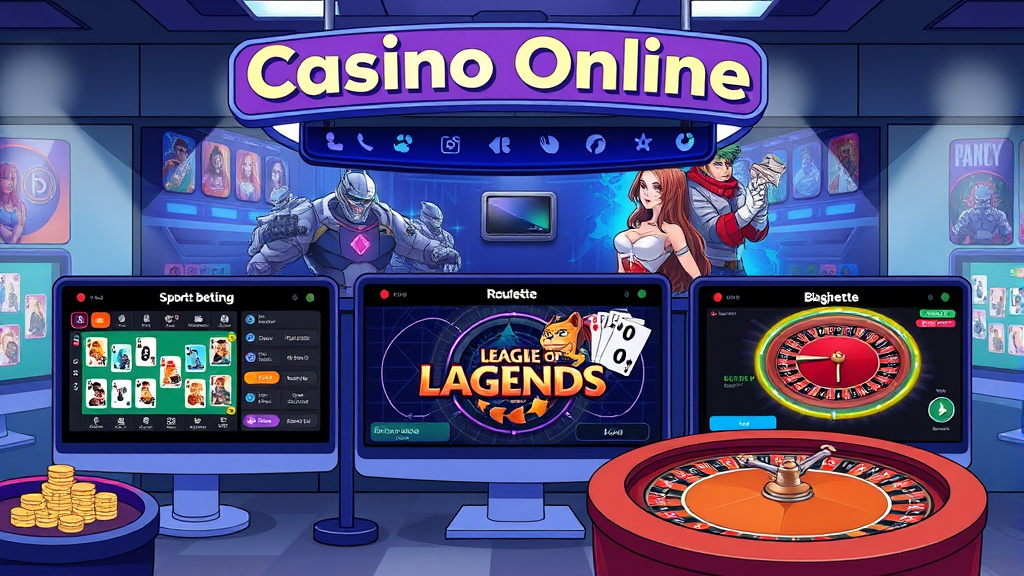
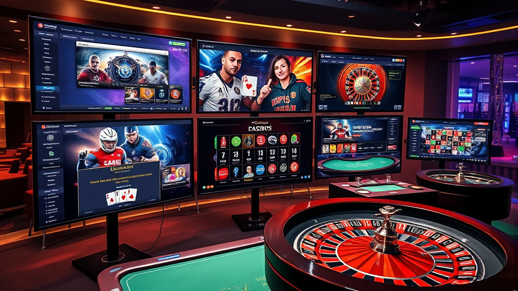
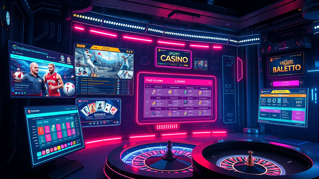 Hình minh họa: SHBET – NỔ HŨ SHBET
Hướng dẫn chi tiết cách thức nhận Freespin hàng tuần tại SHBET
Đầu tiên, để bắt đầu nhận Freespin hằng tuần, bạn cần đăng ký và đăng nhập vào tài khoản SHBET một cách đầy đủ và chính xác. Việc đăng ký rất đơn giản, chỉ cần cung cấp các thông tin cần thiết theo yêu cầu của nhà cái và xác nhận tài khoản qua email hoặc số điện thoại. Sau đó, người chơi cần tích cực tham gia các hoạt động trên nền tảng, như đăng nhập hàng ngày, hoàn thành nhiệm vụ hoặc thắng các trận game Nổ Hũ nhất định để tích lũy điểm thưởng.
Tiếp theo, trong mỗi tuần, nhà cái sẽ tự động gửi thông báo hoặc cập nhật chương trình Freespin về tài khoản của bạn thông qua hệ thống tin nhắn hoặc email. Người chơi chỉ cần đăng nhập vào tài khoản và kiểm tra các phần thưởng nhận được, sau đó kích hoạt Freespin dựa trên hướng dẫn cụ thể của hệ thống. Một số nhà cái yêu cầu nhập mã thưởng hoặc xác nhận qua giao diện người dùng, trong khi số khác sẽ tự động cấp lượt chơi miễn phí cho người chơi đủ điều kiện. Quan trọng nhất là bạn cần chú ý các mốc thời gian đăng ký và hoàn thành các nhiệm vụ để không bỏ lỡ các ưu đãi hấp dẫn này.
Cuối cùng, sau khi nhận Freespin, bạn hãy tận dụng tối đa các lượt quay này để đánh giá các game Nổ Hũ yêu thích, lựa chọn các chiến thuật phù hợp để thắng lớn. Đặc biệt, hãy theo dõi các khuyến mãi đi kèm hoặc các sự kiện đặc biệt dành cho thành viên tích cực nhằm tối ưu hóa khả năng chuyển đổi Freespin thành các khoản thưởng thực tế. Điều này đòi hỏi sự kiên nhẫn, tính toán và nắm vững quy trình của nhà cái, từ đó giúp bạn có thể duy trì nguồn lợi nhuận ổn định trong quá trình chơi.

Điều kiện và điều khoản áp dụng cho chương trình Freespin Nổ Hũ tại SHBET
Mặc dù chương trình Freespin mang lại nhiều lợi ích, nhưng người chơi cần chú ý đến các điều kiện và điều khoản được quy định rõ ràng để tránh các rắc rối hay hiểu lầm không cần thiết. Thứ nhất, mỗi người chơi chỉ được nhận một số lượng Freespin nhất định theo tuần hoặc theo hạn mức của từng chương trình, nên việc quỹ dành cho mỗi người là có giới hạn rõ ràng.
Ngoài ra, các Freespin thường đi kèm với yêu cầu cược tối thiểu hoặc giới hạn trong việc áp dụng cho các game nổ hũ cụ thể, đồng thời cũng có thời hạn sử dụng riêng. Người chơi phải sử dụng đúng hạn, nếu không sẽ bị mất quyền lợi. Các điều kiện về độ tuổi, quốc tịch, hoặc các giấy tờ xác minh danh tính cũng cần được đáp ứng để đảm bảo tính hợp lệ của phần thưởng. Do đó, bạn nên dành thời gian đọc kỹ các điều khoản trong phần chính sách khuyến mãi của SHBET để chắc chắn mình tuân thủ và tối đa hóa lợi ích từ các ưu đãi này.
Thêm vào đó, việc tham gia các trò chơi khác ngoài game Nổ Hũ hoặc cố tình vi phạm điều khoản về gian lận, thao tác bot sẽ dẫn đến việc bị khóa tài khoản hoặc hủy bỏ toàn bộ phần thưởng. Chính vì vậy, người chơi cần trung thực, chơi có trách nhiệm và tuân thủ mọi quy định nhằm bảo vệ quyền lợi cá nhân đồng thời góp phần xây dựng cộng đồng chơi game lành mạnh, minh bạch. Tuân thủ những điều khoản này chính là yếu tố quyết định giúp bạn giữ vững cơ hội nhận thưởng lâu dài từ chương trình Freespin của SHBET.
Lợi ích khi nhận Freespin hàng tuần khi chơi Nổ Hũ tại SHBET
Chương trình Freespin hàng tuần mở ra một loạt lợi ích rõ ràng và thiết thực cho người chơi, giúp nâng cao trải nghiệm chơi game một cách đáng kể. Đầu tiên, việc nhận Freespin giúp giảm thiểu rủi ro về tài chính khi chơi game Nổ Hũ, đặc biệt là với các người chơi mới hoặc những người hạn chế về ngân sách. Bạn có thể thử vận may, rút kinh nghiệm và khám phá các chiến thuật chơi mà không cần phải bỏ ra quá nhiều tiền thật, từ đó gia tăng khả năng thắng lớn trong những vòng chơi tiếp theo.
Thứ hai, Freespin còn mang lại cơ hội tích lũy phần thưởng lớn hơn, giúp người chơi nâng cao tỷ lệ chiến thắng và đạt jackpot dễ dàng hơn. Chương trình này còn tạo điều kiện để bạn thử sức trong các game mới hoặc các phiên bản khác nhau của game Nổ Hũ mà không cần lo lắng về chi phí, từ đó mở rộng kiến thức và kỹ năng chơi game của mình. Ngoài ra, những phần thưởng này còn kết hợp nhiều ưu đãi đặc biệt đi kèm như thưởng tích lũy, vòng quay thưởng thêm hay các phần quà khác, giúp bạn có cảm giác thư giãn, thoải mái hơn khi tham gia thế giới game đổi thưởng trực tuyến.
Không những thế, nhận Freespin hàng tuần còn giúp xây dựng thói quen chơi đều đặn, liên tục và có trách nhiệm. Người chơi sẽ tự rèn luyện khả năng quản lý ngân sách, nâng cao khả năng phân tích và ra quyết định đúng đắn dựa trên các lượt quay miễn phí. Từ đó, việc chơi game không chỉ còn là giải trí mà còn phù hợp với mục tiêu kiếm thưởng hợp pháp, chiến thắng bền vững lâu dài qua từng vòng chơi. Chương trình này chính là cầu nối hoàn hảo để góp phần làm đa dạng trải nghiệm chơi game của bạn, biến những lượt quay vô nghĩa thành những cơ hội thắng lợi thật sự.
Các loại game Nổ Hũ áp dụng chương trình Freespin tại SHBET
SHBET cung cấp đa dạng các loại game Nổ Hũ phù hợp với sở thích, phong cách và chiến lược chơi của từng người chơi. Trong đó, các game slot truyền thống, game chơi theo bộ hình như Cleopatra, Pharaoh’s Gold, hay các phiên bản như Megaways cũng đều nằm trong danh sách áp dụng Freespin, giúp người chơi có nhiều lựa chọn để thử vận may của mình. Chính sự đa dạng này giúp nâng cao tính linh hoạt trong chiến thuật và làm cho trải nghiệm game trở nên hấp dẫn hơn bao giờ hết.
Bên cạnh đó, các dạng game Nổ Hũ hiện đại tích hợp công nghệ đồ họa sống động, thưởng thức âm thanh chân thực cùng các tính năng đặc biệt như wild, scatter, bonus game, free spin nhiều cấp độ đều được nhà cái hỗ trợ tối đa trong chương trình Freespin. Điều này giúp người chơi không bị giới hạn về thể loại, từ đó phần thưởng có thể đa dạng hơn từ các vòng quay miễn phí này. Ngoài ra, các game đổi thưởng có tỉ lệ trả thưởng cao cũng nằm trong danh mục áp dụng Freespin, giúp tăng khả năng thắng lớn và tối đa lợi ích cho người chơi.
Chơi các phiên bản game đa dạng còn giúp người chơi nâng cao kỹ năng, thử nghiệm các chiến thuật khác nhau để tối đa hóa số tiền thắng từ Freespin. Từ đó, bạn có thể dễ dàng nhận diện các game phù hợp nhất với khả năng và sở thích của mình, tiết kiệm thời gian và công sức. Phương thức này còn giúp nhà cái nâng cao chất lượng dịch vụ, giữ chân khách hàng lâu dài hơn qua những trải nghiệm đa dạng, hấp dẫn. Tham gia chơi mọi loại game hot, hot nhất hiện nay trong danh mục Nổ Hũ chính là chìa khóa để biến các lượt Freespin thành các phần thưởng thực sự giá trị.
Mẹo và thủ thuật tối ưu hóa việc sử dụng Freespin để tăng cơ hội thắng
Việc sử dụng Freespin một cách thông minh chính là yếu tố quyết định giúp bạn tối đa hóa phần thưởng từ các vòng quay miễn phí này. Đầu tiên, hãy lựa chọn các game có tỷ lệ trả thưởng cao hoặc có khả năng kích hoạt các chế độ thưởng đặc biệt như Bonus hay Free Spin nhiều cấp độ. Các trò chơi này thường sẽ giúp bạn gia tăng số điểm, thưởng thêm nhiều vòng quay hoặc jackpot lớn hơn khi kết hợp đúng chiến thuật. Kỹ năng đọc hiểu về các biểu tượng, quy luật, cũng là mẹo vặt quan trọng để tối ưu hóa tỉ lệ thắng lợi từ Freespin.
Thứ hai, hãy chơi một cách kiên nhẫn và có kế hoạch rõ ràng. Đặt ra giới hạn thắng thua hợp lý để tránh rủi ro mất hết khi chơi quá lâu hoặc bị ảnh hưởng cảm xúc. Chia những lượt Freespin thành các chu kỳ nhỏ, tập trung vào các chiến thuật đã chuẩn bị sẵn, như tăng cược dần dần hoặc chọn các dòng thắng có xác suất cao, sẽ giúp bạn kiểm soát tốt hơn quỹ chơi của mình. Ngoài ra, việc tham khảo các mẹo chơi từ cộng đồng, các bài viết chia sẻ kinh nghiệm hoặc cập nhật trend mới sẽ giúp bạn không bỏ lỡ các thủ thuật có thể nâng cao khả năng thắng lớn khi sử dụng Freespin.
Cuối cùng, đừng quên tận dụng các chương trình khuyến mãi đi kèm như thưởng sinh nhật, thưởng theo lượt tích lũy hoặc các sự kiện đặc biệt tổ chức trong tuần để cộng dồn lượng Freespin hoặc mở rộng cơ hội thắng lớn hơn. Theo dõi thường xuyên các cập nhật của nhà cái shim, hệ thống sẽ giúp bạn có chiến thuật phù hợp và linh hoạt hơn trong việc sử dụng các lượt quay miễn phí này. Với kế hoạch rõ ràng, mẹo chơi hợp lý, bạn hoàn toàn có thể biến các lượt Freespin thành chìa khóa dẫn đến chiến thắng lớn, đẩy lợi nhuận của mình lên mức cao nhất.
So sánh chương trình Freespin của SHBET với các nhà cái khác
Chương trình Freespin của SHBET nổi bật vì sự linh hoạt, tính công bằng và đa dạng trong các ưu đãi dành cho người chơi. Thông thường, các nhà cái khác cũng có chương trình khuyến mãi Freespin, nhưng mức độ cạnh tranh và điều kiện từ họ vẫn khá khác biệt. Một điểm mạnh của SHBET là khả năng cá nhân hóa các lượt Freespin theo thành tích hoặc hoạt động của từng người chơi, giúp họ cảm thấy được quan tâm và ưu đãi hơn trong hành trình trải nghiệm game.
Ngoài ra, nhà cái này còn có chính sách minh bạch rõ ràng, không gian xử lý và cấp phát thưởng nhanh chóng, dễ hiểu, hạn chế tối đa các rắc rối phát sinh về luật chơi hoặc điều khoản không rõ ràng. Trong khi đó, các đối thủ cạnh tranh thường có giới hạn về thể loại game áp dụng, hoặc quy định thời gian sử dụng Freespin khá hạn chế. SHBET còn thường xuyên cập nhật các chương trình mới, mang đến nhiều hơn các lợi ích đặc biệt phù hợp với xu hướng chơi game hiện đại, sáng tạo hơn.
Về cơ bản, so sánh giữa các nhà cái, điều làm nên sự khác biệt lớn chính nằm ở mức độ linh hoạt, tính minh bạch và sự đổi mới của chương trình Freespin. SHBET nổi bật khi cung cấp các ưu đãi theo tuần, khuyến mãi đa dạng, phù hợp nhiều nhóm người chơi, trong khi các nhà cái khác thường chỉ giới hạn trong các chiến dịch ngắn hạn hoặc chương trình có ít sáng tạo. Vì vậy, nếu bạn đang tìm kiếm môi trường chơi game lâu dài, uy tín và đa dạng, SHBET chính là lựa chọn lý tưởng.
Giải đáp các câu hỏi thường gặp về chương trình Freespin Nổ Hũ tại SHBET
Chắc hẳn có nhiều người chơi mới hoặc thắc mắc về các chi tiết cụ thể liên quan đến chương trình Freespin tại SHBET. Một trong những câu hỏi phổ biến nhất chính là “Làm thế nào để biết khi nào tôi nhận được Freespin hàng tuần?”. Thường thì nhà cái sẽ gửi thông báo qua email hoặc hiển thị trong tài khoản, đồng thời chương trình cũng thường xuyên cập nhật trong phần khuyến mãi của nền tảng.
Ngoài ra, nhiều người còn thắc mắc về quy trình chuyển đổi Freespin thành phần thưởng thực tế. Với các quy định của SHBET, các lượt Freespin thường phải đáp ứng yêu cầu cược tối thiểu và có giới hạn thời gian sử dụng rõ ràng. Đặc biệt, vấn đề về giới hạn cược hay các điều kiện phụ khác cũng là câu hỏi thường gặp, và nhà cái luôn cam kết cung cấp rõ ràng các chi tiết này để mọi người yên tâm tham gia. Các câu hỏi liên quan đến điều kiện đăng ký, kiểm tra trạng thái thưởng, hoặc rủi ro mất phần thưởng vì vi phạm luật chơi cũng luôn được giải thích minh bạch để đảm bảo quyền lợi người chơi.
Bên cạnh đó, có những thắc mắc về các game phù hợp, các chiến thuật tối ưu, hoặc làm thế nào để nâng cao tỷ lệ thắng khi sử dụng Freespin. Câu trả lời luôn hướng tới việc người chơi cần tìm hiểu kỹ luật chơi, có kế hoạch rõ ràng, và chơi có trách nhiệm. Trong mọi trường hợp, SHBET luôn cam kết hỗ trợ và cung cấp thông tin chính xác, giúp người chơi dễ dàng nắm bắt mọi chính sách và tối đa hóa lợi ích của mình trong chương trình ưu đãi này.
Lưu ý quan trọng khi tham gia chương trình Freespin để tránh rủi ro
Dù chương trình Freespin mang lại nhiều lợi ích, người chơi cần có ý thức rõ ràng về những lưu ý để tránh các rủi ro không mong muốn. Đầu tiên, bạn nên kiểm tra kỹ các điều kiện và điều khoản của chương trình để không vô tình vi phạm các quy định về độ tuổi, khu vực hoặc hành vi gian lận. Việc này giúp đảm bảo tài khoản của bạn không bị khóa hoặc mất quyền lợi do sơ suất nhỏ.
Tiếp theo, hãy quản lý ngân sách chơi hợp lý, đừng để cảm xúc chi phối quyết định chơi game. Freespin là cơ hội để thử vận may, chứ không phải phương pháp đảm bảo thắng thắng chắc, vì vậy không nên lao vào chơi quá nhiều lượt quay hoặc đặt cược cao bất hợp lý sẽ dễ gây thua lỗ. Luôn giữ thái độ tỉnh táo, biết dừng đúng lúc khi thắng hoặc thua để bảo vệ tài sản của chính mình. Ngoài ra, cần tránh các hành vi gian lận như dùng phần mềm, thao túng hệ thống hoặc chat chít để giành lợi thế không chính đáng, vì hành vi này có thể dẫn đến hậu quả nghiêm trọng.
Một điểm nữa đó là các rủi ro về an ninh tài khoản: bạn cần bảo vệ mật khẩu, không chia sẻ thông tin cá nhân hoặc tài khoản của mình với người khác để tránh bị tấn công hoặc mất mát. Cuối cùng, hãy luôn cập nhật các thông báo, chính sách mới của SHBET để nắm rõ các thay đổi hoặc khuyến mãi mới nhất, nhằm tránh bỏ lỡ các cơ hội thưởng và đảm bảo trải nghiệm chơi game an toàn, hiệu quả hơn.
Kết luận: Tại sao nên tham gia chương trình Freespin Nổ Hũ hàng tuần của SHBET | shbetvy.com
Chương trình Freespin hàng tuần của SHBET thực sự là một cơ hội vàng cho những ai đam mê chơi game Nổ Hũ, muốn tối ưu hóa lợi ích và giảm thiểu rủi ro trong quá trình chơi. Không chỉ đơn thuần là phần thưởng miễn phí, hệ thống này còn giúp bạn nâng cao kỹ năng, thử chiến thuật mới, và trải nghiệm các trận game đầy kịch tính mà không lo ngại về chi phí ban đầu.
Ngoài ra, SHBET còn nổi bật với chính sách minh bạch, dịch vụ hỗ trợ tận tình và các chương trình khuyến mãi đa dạng, phù hợp nhiều nhóm đối tượng. Việc hiểu rõ cách nhận Freespin, điều kiện áp dụng và có những mẹo chơi phù hợp sẽ giúp bạn dễ dàng giành thắng lợi lớn hơn mỗi tuần. Chính vì thế, nếu bạn thực sự đam mê thể loại game Nổ Hũ, hãy đừng bỏ lỡ cơ hội tham gia chương trình ưu đãi hữu ích này để biến các lượt quay miễn phí thành cơ hội chiến thắng thật sự và nâng cao trải nghiệm chơi game của chính mình tại SHBET!

 Hình minh họa: 123bet – nạp tiền 123Bet an toàn và tiện lợi

 Hình minh họa: https://gamebaidoithuong.wales/ – game bài đổi thưởng
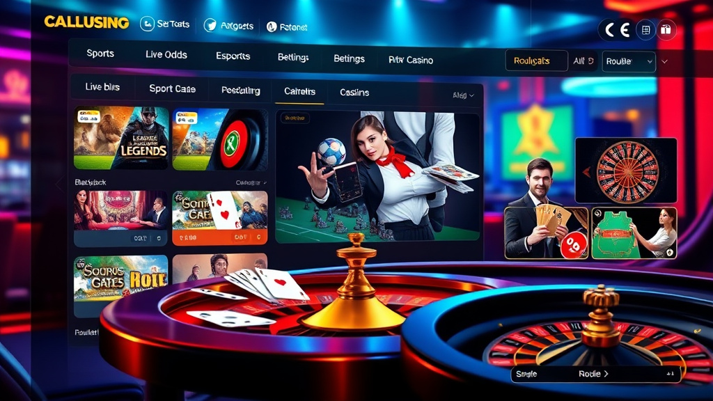

 Hình minh họa: https://hitclubapp.com/ – Hit club

 Hình minh họa: https://hitclub.sbs/
 Hình minh họa: https://sunwin86.page/ – sun win

 Hình minh họa: https://sunwinuk.com/ – Tải Sunwin


 Hình minh họa: https://go88.deal/